Back علم الأحياء البنيوي Arabic Структурна биология Bulgarian Strukturna biologija BS Biologia estructural Catalan Strukturní biologie Czech Strukturbiologie German Struktura biologio Esperanto Biología estructural Spanish زیستشناسی ساختاری Persian Rakennebiologia Finnish
| Part of a series on |
| Biochemistry |
|---|
 |
Structural biology, as defined by the Journal of Structural Biology, deals with structural analysis of living material (formed, composed of, and/or maintained and refined by living cells) at every level of organization. Early structural biologists throughout the 19th and early 20th centuries were primarily only able to study structures to the limit of the naked eye's visual acuity and through magnifying glasses and light microscopes.
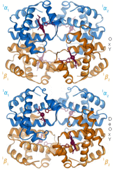
In the 20th century, a variety of experimental techniques were developed to examine the 3D structures of biological molecules. The most prominent techniques are X-ray crystallography, nuclear magnetic resonance, and electron microscopy. Through the discovery of X-rays and its applications to protein crystals, structural biology was revolutionized, as now scientists could obtain the three-dimensional structures of biological molecules in atomic detail.[1] Likewise, NMR spectroscopy allowed information about protein structure and dynamics to be obtained.[2] Finally, in the 21st century, electron microscopy also saw a drastic revolution with the development of more coherent electron sources, aberration correction for electron microscopes, and reconstruction software that enabled the successful implementation of high resolution cryo-electron microscopy, thereby permitting the study of individual proteins and molecular complexes in three-dimensions at angstrom resolution.
With the development of these three techniques, the field of structural biology expanded and also became a branch of molecular biology, biochemistry, and biophysics concerned with the molecular structure of biological macromolecules (especially proteins, made up of amino acids, RNA or DNA, made up of nucleotides, and membranes, made up of lipids), how they acquire the structures they have, and how alterations in their structures affect their function.[3] This subject is of great interest to biologists because macromolecules carry out most of the functions of cells, and it is only by coiling into specific three-dimensional shapes that they are able to perform these functions. This architecture, the "tertiary structure" of molecules, depends in a complicated way on each molecule's basic composition, or "primary structure." At lower resolutions, tools such as FIB-SEM tomography have allowed for greater understanding of cells and their organelles in 3-dimensions, and how each hierarchical level of various extracellular matrices contributes to function (for example in bone). In the past few years it has also become possible to predict highly accurate physical molecular models to complement the experimental study of biological structures.[4] Computational techniques such as molecular dynamics simulations can be used in conjunction with empirical structure determination strategies to extend and study protein structure, conformation and function.[5]


- ^ Curry, Stephen (2015-07-03). "Structural Biology: A Century-long Journey into an Unseen World". Interdisciplinary Science Reviews. 40 (3): 308–328. Bibcode:2015ISRv...40..308C. doi:10.1179/0308018815Z.000000000120. ISSN 0308-0188. PMC 4697198. PMID 26740732.
- ^ Campbell, Iain D. (2013-01-01). "The evolution of protein NMR". Biomedical Spectroscopy and Imaging. 2 (4): 245–264. doi:10.3233/BSI-130055. ISSN 2212-8794.
- ^ Banaszak LJ (2000). Foundations of Structural Biology. Burlington: Elsevier. ISBN 9780080521848.
- ^ Cite error: The named reference
Stoddartwas invoked but never defined (see the help page). - ^ Karplus M, McCammon JA (September 2002). "Molecular dynamics simulations of biomolecules". Nature Structural Biology. 9 (9): 646–652. doi:10.1038/nsb0902-646. PMID 12198485. S2CID 590640.
© MMXXIII Rich X Search. We shall prevail. All rights reserved. Rich X Search